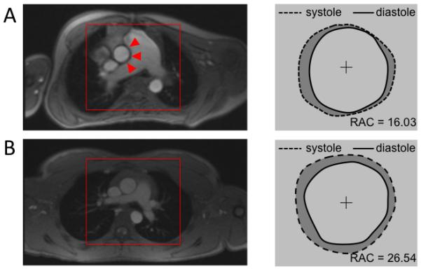
Figure 7.

Pulmonary Artery Impingement Upon Aorta. A) On this representative pediatric PAH patient one can immediately notice a severely dilated MPA impinging on the ascending aorta. Wall deformation analysis from segmented PC-MRI magnitude images shows reduced range of motion along the aortic – MPA interface highlighted by red triangles. B) Conversely, representative control subject reveals size proportional vessel and uniform aortic expansion along the central axis.
