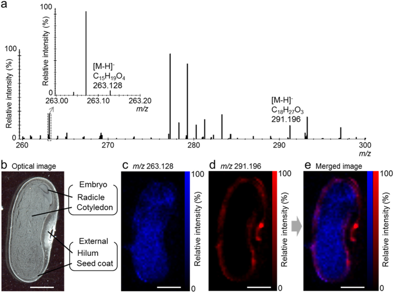
Figure 2. Desorption electrospray ionisation-imaging mass spectrometry (DESI-IMS) of immature Phaseolus vulgaris L. seed.
Sections were prepared from a 163.7-mg seed. Spatial resolution was set at 200 μm. Internal mass calibration was performed post-acquisition using the exact m/z of the palmitic acid [M-H]− ion (m/z 255.233). (a) Mass spectrum at the m/z 260–300. The chemical formulae are shown at the top of the respective peaks. (b) Optical image of a seed section after DESI-IMS. (c) Ion image at m/z 263.128. (d) Ion image at m/z 291.196. (e) Merged ion image of (c and d). Scale bar = 2 mm.

