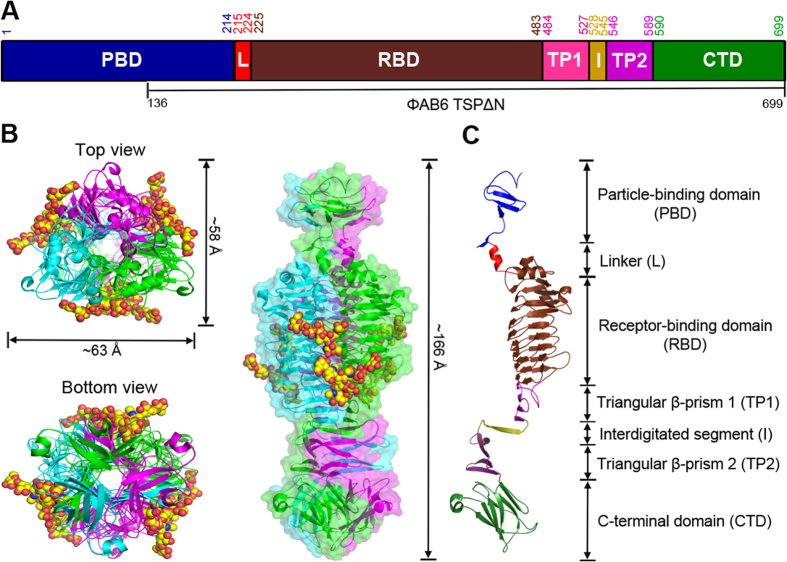
Figure 3. Overall structure.
(A) Domains in full-length ΦAB6 TSP are shown in a schematic representation. The numberings for amino acid residues at the start and end of each domain are further indicated. TSP∆N includes residues 136–699. (B) The TSP∆N trimer is shown as three-colored ribbons in three different views, with bound oligosaccharides shown as space-filled models. The view on the right is perpendicular to the trimer axis onto the intersubunit groove with the N-terminal end on top, and a transparent surface of the structure is shown as well. (C) The seven domains in a TSP∆N monomer are colored as in (A).

