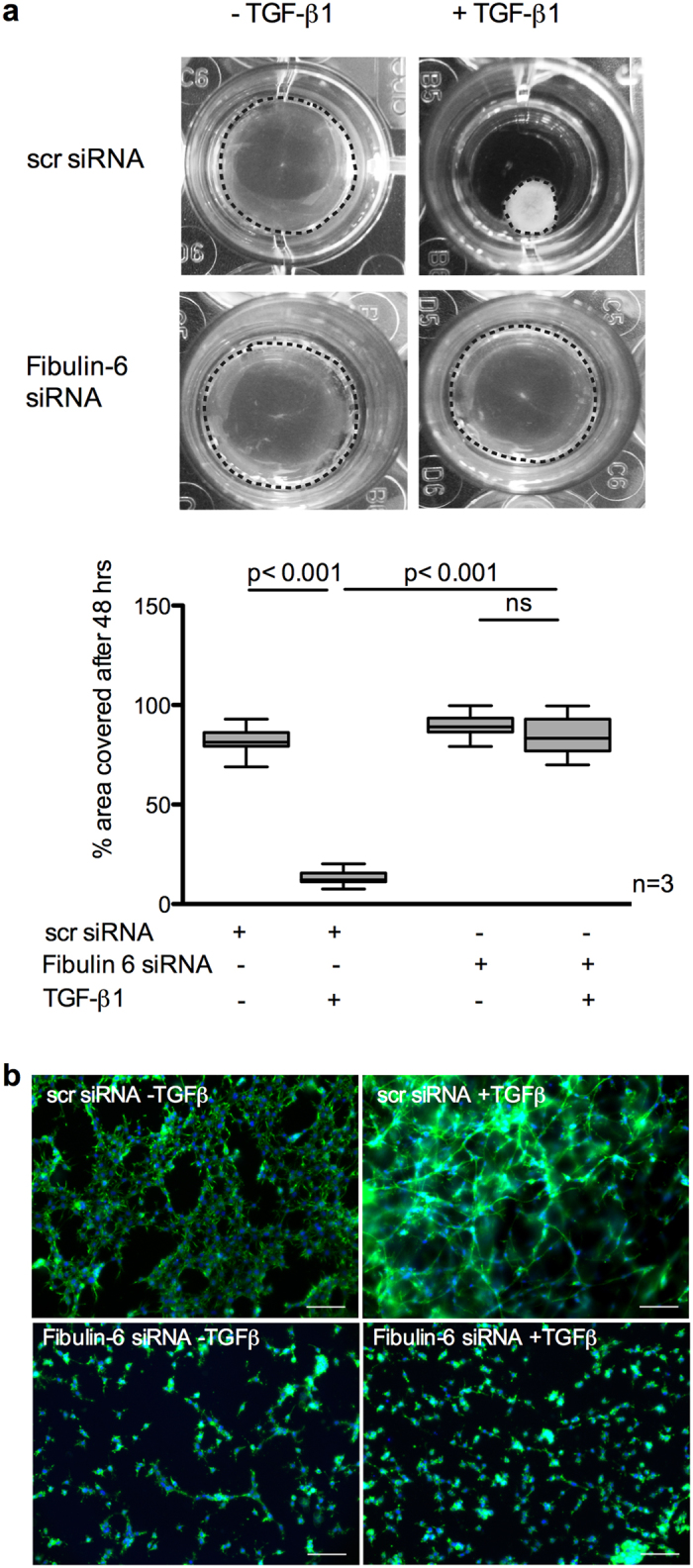
Figure 2. TGF-β mediated collagen contraction is reduced in fibulin-6 KD CF.

Collagen gels carrying either scr-siRNA or fibulin-6 KD 3T3 CF were exposed to TGF-β to induce contraction. (a) A representative picture of collagen gels 48 h after TGF-β stimulation is shown. The area of the gels (marked by the dotted circle around the collagen gel) before and after stimulation was measured using Image J and is represented in the corresponding graph. After TGF-β stimulation the size of collagen gels carrying control cells is significantly reduced while collagen gels carrying fibulin-6 KD cells display no contraction (n = 3, p < 0.001, nonparametric Mann-Whitney U test). The experiment was repeated three times in quadruplicates. (b) Photographs of collagen gels stained for phalloidin after 48 hours of TGF-β stimulation (scale bar = 100 μm). In scr-siRNA transfected 3T3 CF the formation of cellular networks during contraction can be observed, while actin cytoskeletal network and cell-cell contact in fibulin-KD CF are absent.
