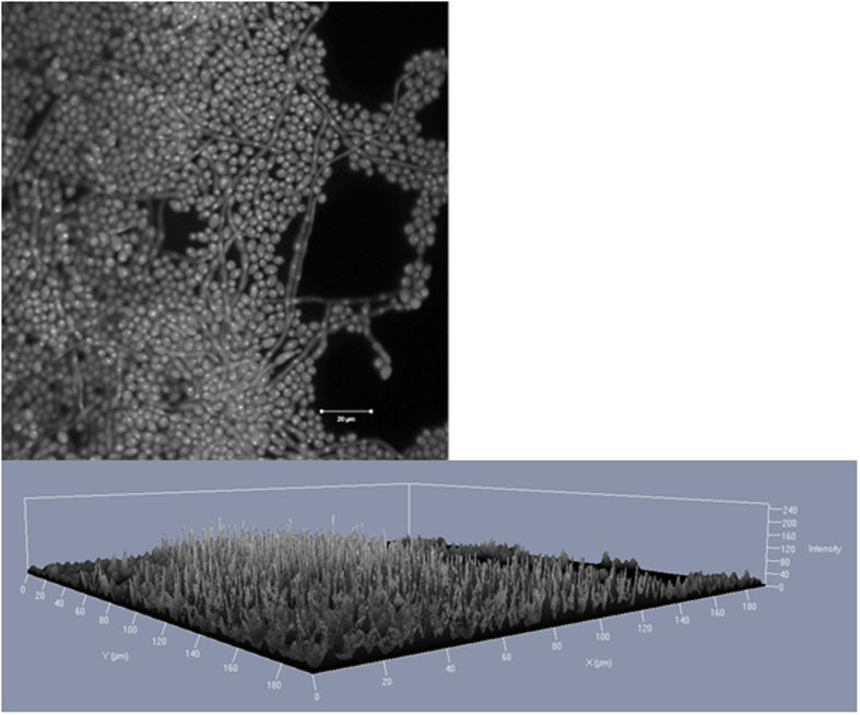
Figure 3. Confocal laser scan microscopy images of C. albicans (ATCC 90028) biofilm grown for 48 hours at 35 °C.
The DNA of the cells was stained by 0.01% acridine orange for 2 minutes. A laser with a wavelength of 488 nm was used. (A) Matured biofilm in a 2D image. Scale bar equals 50 μm. (B) Matured biofilm a 2.5 D image.

