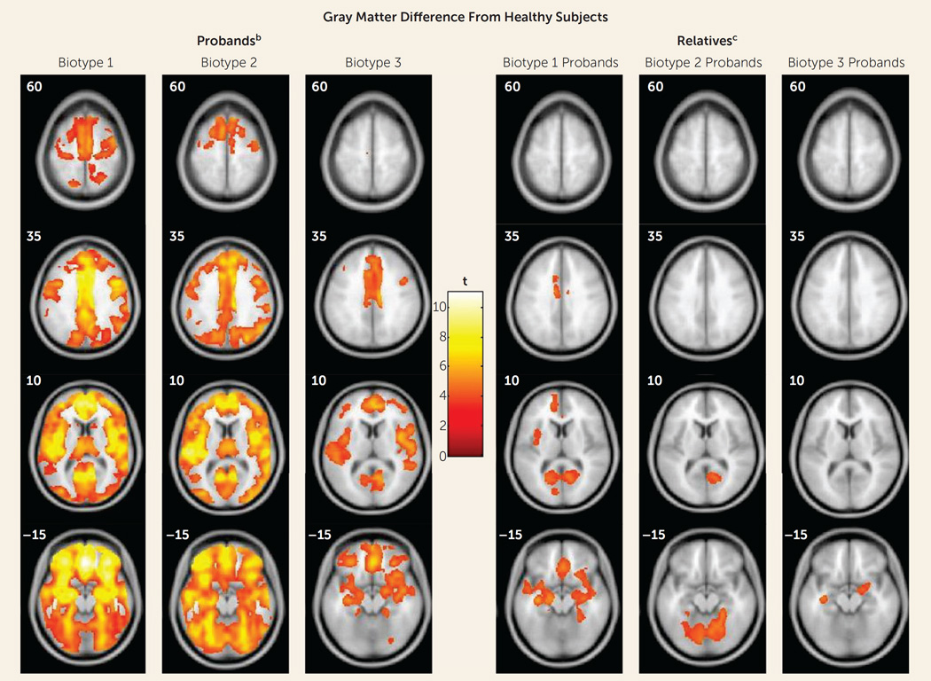
FIGURE 3. Gray Matter Differences From Healthy Subjects in Voxel-Based Morphometry Results for Probands With Psychosis and Their First-Degree Relatives, by Proband Biotypea.
a Images are displayed in neurological convention. Outcomes are reported at p=0.05, with cluster-wise family-wise-error correction.
b Biotype1had the most extensive volume reductions, with the largest effects in the frontal, cingulate, temporal, and parietal cortex, as well as basal ganglia and thalamus. Biotype 2 had volume reductions regionally overlapping with those in biotype 1, with the largest effects in the frontotemporal cortex, parietal cortex, and cerebellum, albeit of a lesser magnitude overall than for biotype 1. Biotype 3 had smaller clusters of reductions that were primarily distributed over frontal, cingulate, and temporal regions.
c The biological relatives of biotype 1 probands showed predominantly anterior, mostly frontotemporal, gray matter volume differences. The relatives of biotype 2 probands showed posterior, mostly cerebellar, reductions. The relatives of biotype 3 probands showed small clusters of reductions limited to bilateral temporal regions and right inferior frontal regions.

