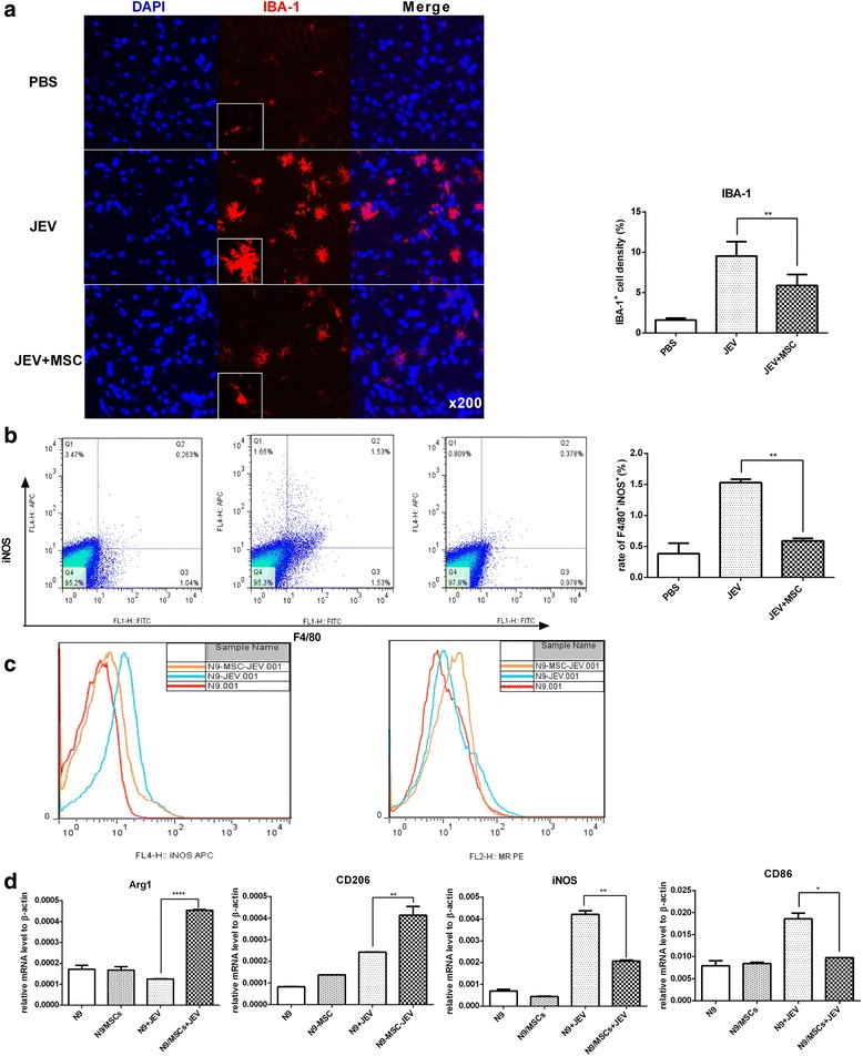
Fig. 3.

MSC transplantation inhibits the activation of microglia and regulates the M1-to-M2-like phenotypic switching. a The activation of microglia was detected by anti-IBA-1 antibody with the boxed areas showing a higher magnification (left), and the intensity of IBA+ cells of each group was analyzed with ImageJ (right) (PBS n= 3, JEV n= 6, JEV + MSCs n= 6; three sections per animal, five fields per section). b Total brain tissue was harvested, and the percentage of F4/80+ iNOS+ cells (left), which represents the M1 polarized macrophages in the brain, was calculated; the mean of the percentage (right) of M1 macrophages from each group was summarized (PBS n= 2, JEV n= 3, JEV + MSC n= 3). c In vitro cultures of the microglia line N9 with or without MSCs (10:1, respectively) were infected with JEV (MOI = 5, 48 h), and the expression levels of the M1 marker iNOS and the M2 marker MR (CD206) were analyzed by Flow Jo. d The expression levels of Arg1 and CD206 (markers for M2) as well as iNOS and CD86 (markers for M1) in N9 and N9-MSC cultures either with or without JEV infected with JEV (MOI = 5, 48 h) was detected by qRT-PCR. The data represent the mean ± SEM for three independent experiments. *P < 0.05, **P < 0.01, ****P < 0.0001. Arg1 arginase-1, IBA-1 ionized calcium binding adaptor molecule-1, iNOS inducible nitric oxide synthase, JEV Japanese encephalitis virus, MSC mesenchymal stem cell, PBS phosphate-buffered saline
