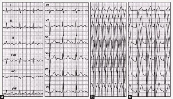
Figure 1.
Atrial arrhythmias during vasodilator stress. Electrocardiogram tracings obtained at baseline (panel a), during atrial flutter (b), and fast atrial fibrillation (2 min after aminophylline, c). Left bundle branch block morphology with no R-wave progression through chest leads denoted counterclockwise rotation of the heart in horizontal plane

