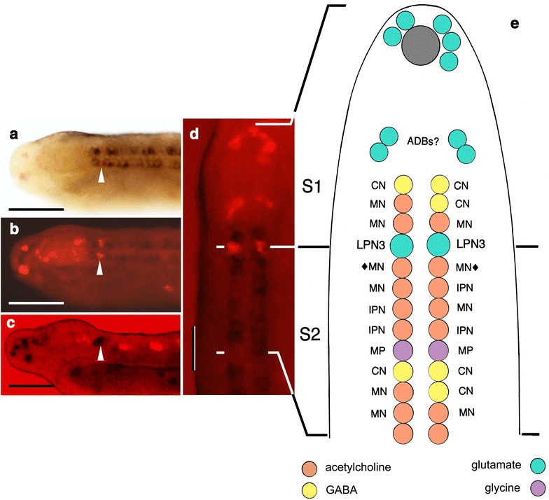
Fig. 2.

In situ data on transmitter localization. a, b Images from Fig. 10g–i in [8], showing VAChT (dark purple) and VGLUT (red) expression, markers for cholinergic and glutamatergic neurons, respectively. c Image from Fig. 9 g in [8], showing VGLUT expression in dark purple and VGAT (GAD+) expression in red, markers for glutamatergic and GABAergic neurons, respectively. Arrowheads in (a–c) indicate the position of the LPN3s; the loose cluster of red cells just forward of this point in (c) are the putative anterior group CNs. Scale bars for (a–c) 50 μm. d A dorsal view of the specimen in (a) reoriented for comparison with the diagram; the junction between somites 1 and 2 corresponds with the position of the LPN3s as shown. Scale bar 5 μm. e Revised neurotransmitter map, an update combining Figs. 3 and 12 from [8] showing the anterior end of the dorsal nerve cord in 20–24 h neurulae of B. floridae to the end of somite 2, opened out and viewed from above, with each of the ventral neurons expressing markers for known transmitters matched with the corresponding cell type from TEM. Neurons: commissural neurons (CN), third pair of large paired neurons (LPN3); motoneurons (MN, diamonds mark the first pair of DC motoneurons), multipolar neurons (MP), ipsilateral projection neurons (IPN). The color code, by transmitter, is shown at the bottom. The figure is altered from the original to show how the anterior-most glutamatergic neurons are positioned at 24 h based on (d). Several such cells cluster around the anterior pigment spot (gray) belonging to the frontal eye, but we are uncertain as to their identity. We interpret the paired dorsolateral groups of cells between the pigment spot and the ventral columns as anterior dorsal bipolar cells (ADBs) as indicated
