Figure 5.
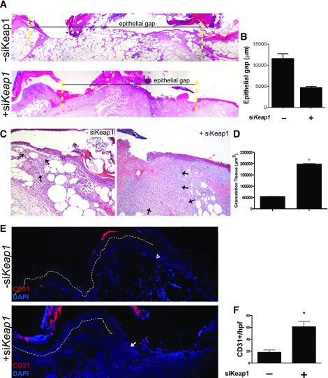
Topical siKeap1 gene therapy improves the histologic profile of diabetic wounds. Ten-day-old diabetic wounds were analyzed histologically to study the effect of topical siKeap1 therapy on reepithelialization and neovascularization. A: Hematoxylin and eosin–stained sections demonstrate accelerated reepithelialization in siKeap1-treated wounds. The yellow dotted lines indicate wound boundaries. B: Quantification of the epithelial gap. C: siKeap1-treated diabetic wounds demonstrate increased granulation tissue area. The black arrows indicate granulation tissue. D: Quantification of granulation tissue. siKeap1 therapy increases granulation tissue production by >3.5-fold compared with scramble-treated controls (*P < 0.01). E: CD31 immunofluorescence on tissue sections demonstrates increased neovascularization within the wound bed in siKeap1-treated wounds. The yellow dotted line demarcates the epidermis (above) and dermis (below). The wound bed is to the right of the yellow dotted line. The open arrowhead indicates low expression; the white arrow indicates high expression. F: Quantification of CD31+ cells in granulation tissue (*P < 0.01).

