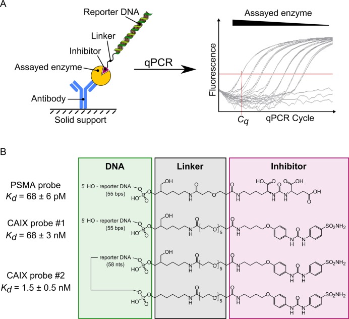
Figure 1.
Schematic representation of enzyme detection using DIANA. (A) A covalent conjugate comprising an oligonucleotide (reporter DNA) and small-molecule competitive inhibitor of the target enzyme is used as a detection probe. The probe binds to the active site of the enzyme, which has been captured on the solid support by binding to the immobilized antibody. The amount of detection probe bound to the target enzyme is measured by quantitative PCR in terms of the threshold cycle (Cq), which is indirectly proportional to the logarithm of probe concentration. (B) Structures of the detection probes used for quantification of PSMA and CAIX. Each probe consists of reporter DNA (green box) covalently attached via a linker region (black box) to a competitive inhibitor of PSMA or CAIX (magenta box). The PSMA probe and CAIX probe #1 contain one copy of inhibitor linked to the 3′ end of the double-stranded reporter DNA, whereas CAIX probe #2 contains two copies of the inhibitor linked to both ends of the single-stranded reporter DNA. Probe affinities (Kd values) were determined by titrating each probe into wells containing the corresponding captured enzyme.

