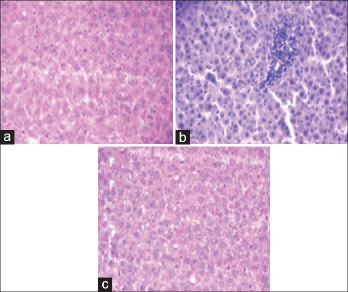
Figure 6.

Photomicrograph of liver tissue (a) control rats showing normal hepatic cells with central vein and Sinusoidal dilation, (b) acetaminophen treated rats showing nonspecific inflammation of hepatic tissue with prominent features of vasculitis (c) rats supplemented with alpha-ketoglutarate along with acetaminophen showing milder degree of inflammation as compared to acetaminophen alone
