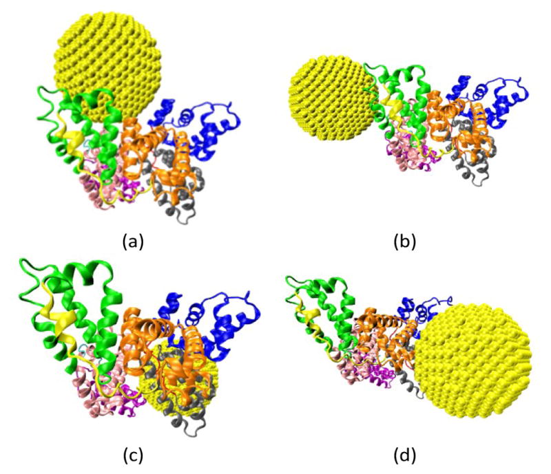
Fig. 3.

Four binding configurations of an HSA protein and a 4.0 nm gold NP. (a) Complex A, (b) Complex B, (c) Complex C and (d) Complex D. The HSA protein is shown in the New Cartoon form and the gold atoms are shown in the VDW form. The subdomains of the HSA are shown in different colors (Subdomain Ia: 5–105, Ia–Ib loop: 106–118, Subdomain Ib: 119–195, Subdomain IIa: 196–292, IIa–IIb loop: 293–313, Subdomain IIb: 314–383, Subdomain IIIa: 384–491, IIIa–IIb loop: 492–509, IIb subdomain: 510–582). The figures are generated using the VMD package49
