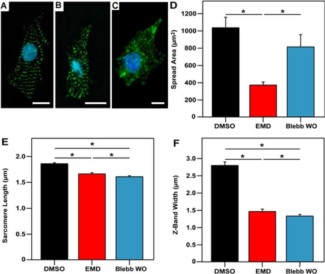
Figure 5.
EMD and blebbistatin impair myofibril structure. Fluorescent images showing nuclei (cyan) and α-actinin (green) for representative cardiomyocytes from the (A) DMSO-treated group, (B) EMD-treated group and (C) blebbistatin-treated. (D) Spread area was found to be significantly less for the EMD (377 ± 31 µm2) as compared to the DMSO (1041 ± 117 µm2) and blebbistatin (819 ± 138 µm2) groups. Measurement of (E) sarcomere lengths (1.87 ± 0.01 µm for DMSO, 1.67 ± 0.02 µm for EMD, and 1.61 ± 0.01 µm for blebbistatin) and (F) Z-band widths (2.81 ± 0.09 µm for DMSO, 1.48 ± 0.06 µm for EMD, and 1.34 ± 0.04 µm for blebbistatin) also show a significant reduction with the addition of EMD or blebbistatin. Scale bar represents 10 µm. Data is from at least 11 cells per group.

