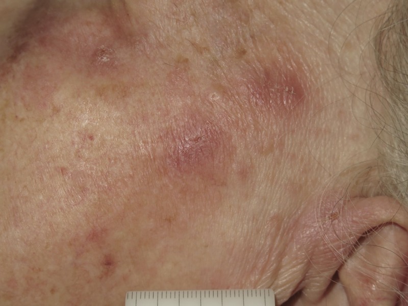
Figure 2.

A close-up clinical photo of one of the lesions, highlighting that the eruption consisted of slightly excoriated papules. The clinical differential diagnosis was broad, including discoid lupus erythematosus, sarcoidosis, lymphoma and pseudolymphoma. [Copyright: ©2017 Lallas et al.]
