Figure 3.
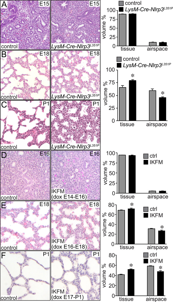
Macrophage activation inhibits late stage airway morphogenesis. (A–C). Abnormal lung morphogenesis in LysM-Cre-NLRP3L351P mice. Lungs from LysM-Cre-NLRP3L351P mice and littermate controls were fixed, processed, and sectioned. Representative images from H&E stained sections from E15 (A), E18 (B) and postnatal day 1 (P1, C) mice are shown. Lung morphometry measurements at right showed no difference in mutant and control lungs at E15, but increased lung interstitial tissue volume and reduced airspace volume in E18 LysM-Cre-NLRP3L351P lungs (*P < 0.01, n = 16). Low viability in P1 LysM-Cre-NLRP3L351Pmice prevented analysis at postnatal time points. (D–F). To measure stage-dependent effects of macrophage activation on lung development, pregnant IKFM mice were provided with drinking water containing doxycycline (2 g/L) for 48 h periods from (D) E14-E16, (E) E16-E18, or (F) E17-P1. Following 48 h of treatment, lungs from IKFM and littermate control embryos were fixed, processed, and imaged for lung morphometry measurements. Representative images are shown. E16 control and IKFM lungs did not have measurable differences in airspace formation. However IKFM lungs contained more simplified airspaces and thicker lung mesenchyme at E18 and P1. (*P < 0.01, n = 94–126.)

