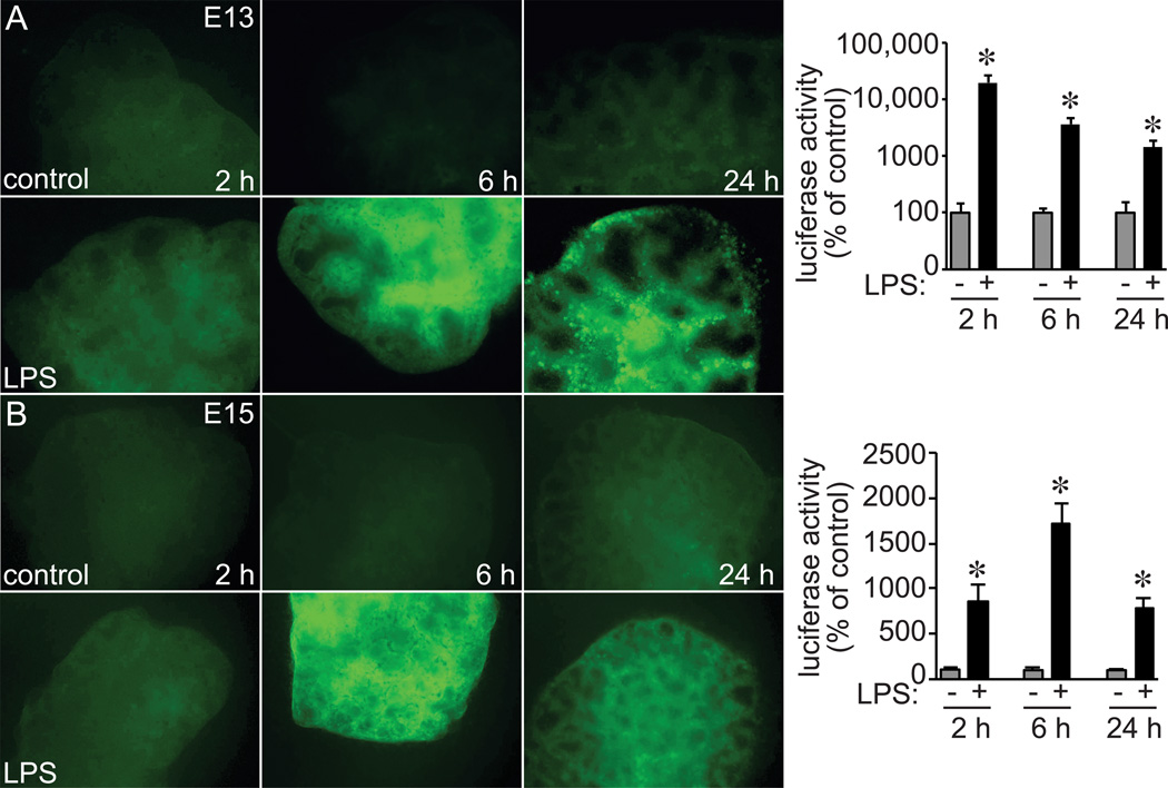
Figure 6.
LPS induced NF-κB activation in early stage fetal mouse lung explants. (A) E13 and (B) E15 lung explants from NGL reporter mice were treated with LPS for up to 24 h. (A,B). GFP reporter expression was imaged by fluorescence microscopy and detected following LPS treatment in both E13 and E15 explants (representative images shown). NF-κB reporter expression was also quantified by luciferase activity in parallel experiments. Each condition and time point normalized to total protein and compared to untreated controls (*P < 0.05, n = 6).

