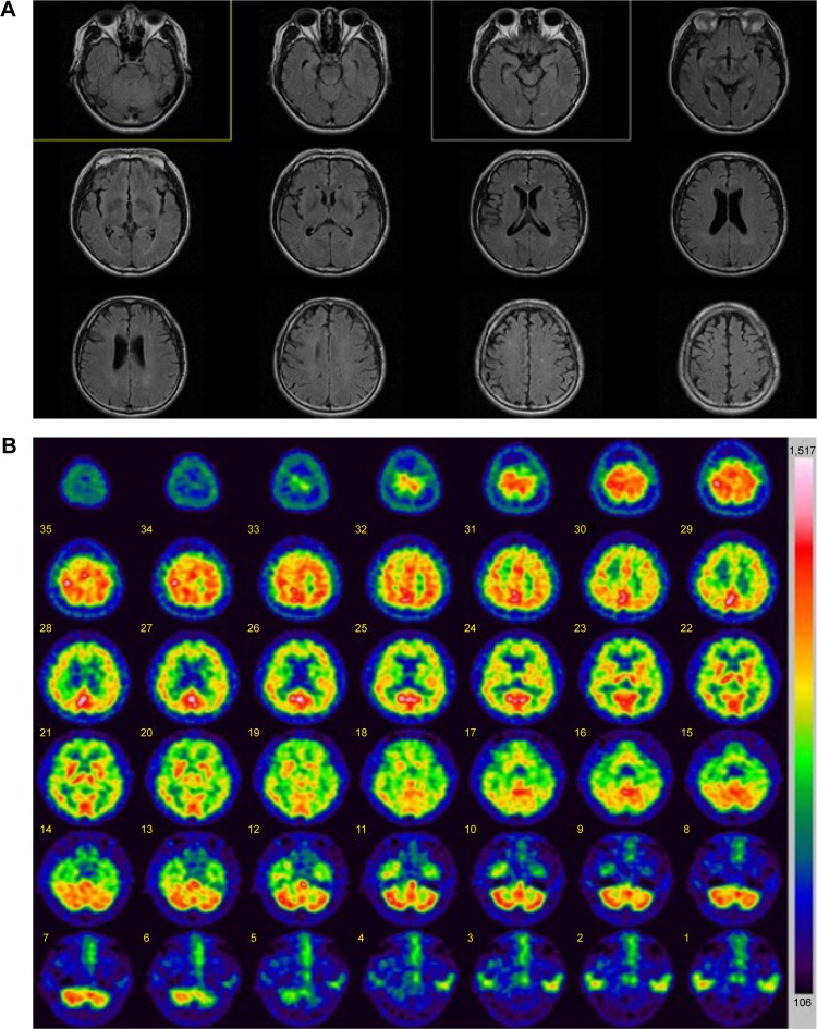
Figure 2.
Imaging data of the patient with PSEN2 R62C.
Notes: (A) MRI data: mild cortical atrophy was present, but without lesions. (B) SPECT data: hypoperfusion was observed in frontal, limbic, and temporal brain areas.
Abbreviations: MRI, magnetic resonance imaging; SPECT, single-photon emission computed tomography.

