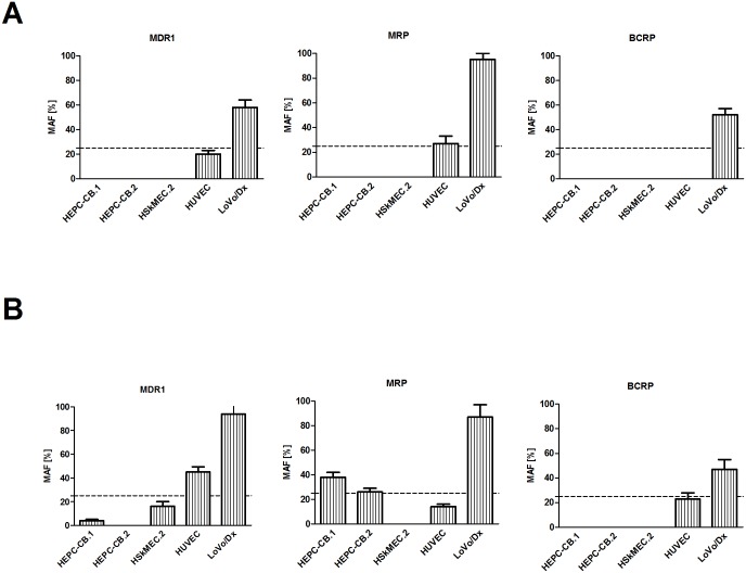
Fig 4. Comparison of MDR activity factor (MAF) of endothelial cells: A) standard functional test; B) commercial functional eFluxx-ID Green test.
A) Cell lines were trypsinized, washed with medium and incubated with rhodamine 123 or calcein AM dyes. After one wash, cells were incubated in medium or medium with specific inhibitors: 10 μM of verapamil, 25 μM of MK-571 or 20 μM of novobiocin. B) Cell lines were trypsinized, washed with PBS, aliquoted and treated in triplicate with different inhibitors (20 μM of verapamil, 25 μM of MK-571, or 50 μM of novobiocin) or untreated (medium with DMSO). Tested probes (eFluxx-ID Green) were added to every sample apart from one tube (white cells). The cells were incubated with the dye in the presence or absence of inhibitors for 30 min. at 37°C.

