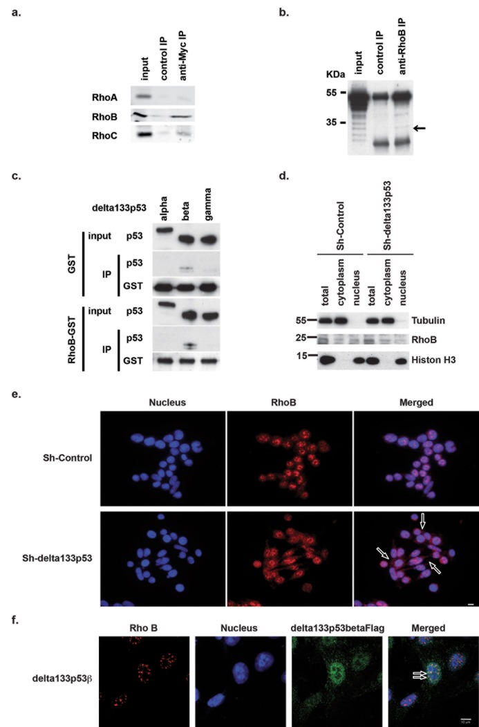
Fig 1. Delta133p53ß physically interacts with RhoB.
(a) Immunoblot analysis showing the specific co-immunoprecipitation of MYC-tagged delta133p53ß and endogenous RhoB or RhoC, to a lower extent, but not RhoA. (b) Immunoblot analysis showing the co-immunoprecipitation of endogenous RhoB and delta133p53ß with anti-RhoB antibodies. Arrow shows co-immunoprecipitation of delta133p53ß isoform. (c) In-vitro binding assay showing the direct interaction between recombinant RhoB-GST fusion protein and delta133p53ß, but not α or γ. (d) Cellular fractionation showing RhoB cytoplasmic re-localization in SW620 cells transfected with shRNAS against delta133p53 compared with shControl (mock-transfected cells). (e) Immunofluorescence analysis of RhoB localization in control SW620 cells (shControl) or after transfection with shdelta133p53. Arrows indicate examples of RhoB cytoplasmic localization. Scale bar: 10μm. (f) Confocal images showing the co-localization of RhoB and delta133p53ß in the nucleus of SW480 cells that overexpress delta133p53ß. Arrows indicate examples co-localization of RhoB and delta133p53ß in the nucleus. Scale bar: 10μm.

