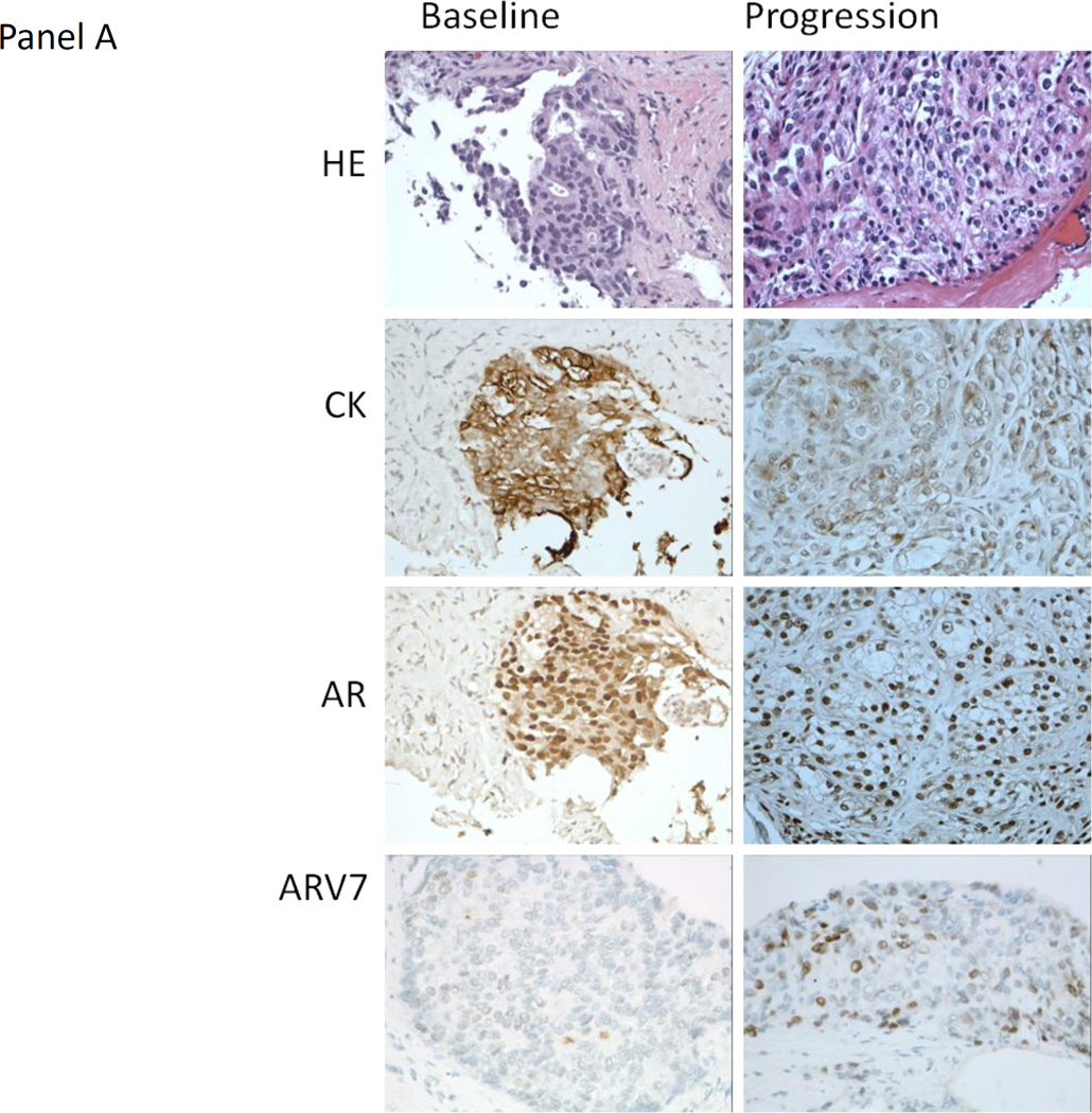
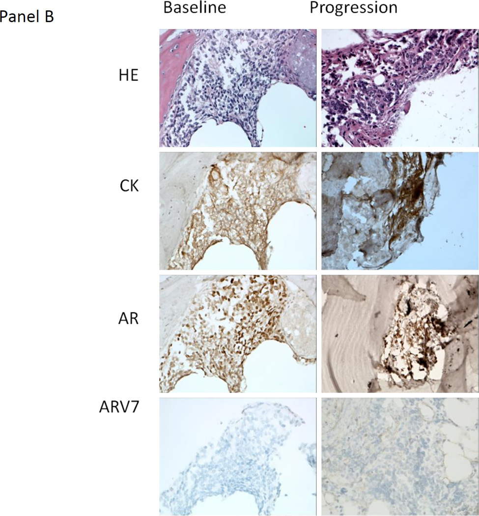
Figure 3.
A) Representative hematoxylin and eosin (HE) staining, and immunohistochemistry staining for cytokeratin (CK), the androgen receptor (AR) and AR variant 7 (ARV7) from the baseline and progression lymph node metastasis biopsy obtained from patient 36 (40X). The HE morphology of the baseline and progression biopsies was similar. ARV7 was weakly staining (+1, <10% of tumor nuclei at given intensity) at baseline and moderate staining (+2, ~50% of tumor nuclei at given intensity) at progression. This patient had lymph node only metastases and remained on treatment for 5.5 months after experiencing disease progression. B) Representative HE staining, and IHC staining for CK, the AR and ARV7 from the baseline and progression bone metastasis biopsy obtained from patient 33 (40X). The HE morphology of the baseline and progression biopsies was similar. There was no ARV7 staining at baseline or progression. The patient had bone only metastases and remained on treatment for 8 months after experiencing disease progression.


