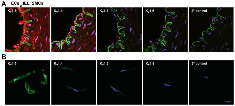
Figure 5. Immunofluorescence localization of KV1 α-subunits in human adipose arterioles.
KV1.5 is the major KV1 channel protein expressed in human adipose arteriolar smooth muscle cells. A: Confocal immunofluorescence images of KV1 α-subunit proteins (red) in cross tissue sections (10 μm) of an intact human adipose arteriole. KV1.5 and 1.4 of a lower level were detected in smooth muscle cells (SMCs) and endothelial cells (ECs). KV1.3 and 1.6 subunits were also faintly visible in ECs. Cell nuclei were stained with DAPI (blue). IEL, internal elastic lamina (green auto-fluorescence). B: Confocal immunofluorescence images of corresponding KV1 α-subunit proteins (green) in freshly dissociated SMCs from a human adipose arteriole. KV1.5 protein was mainly localized on the cell membrane of SMCs. Cell nuclei was stained with DAPI (blue). Data are representative of >3 independent tissues.

