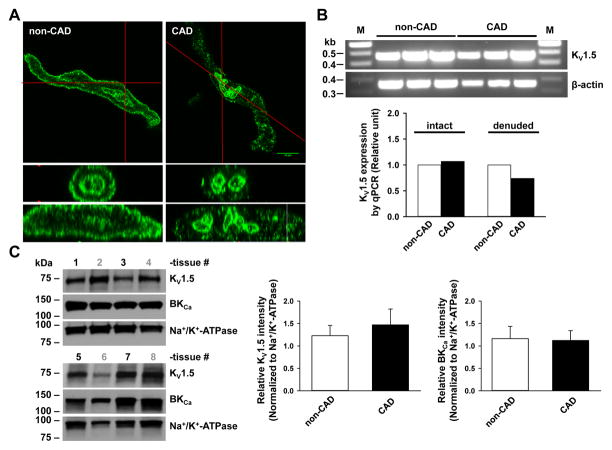
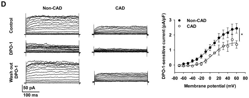
Figure 8. Comparison of KV1.5 subcellular localization, total mRNA and protein expression, and K+ current in non-CAD and CAD human arterioles.
A: Immunofluorescence detection of KV1.5 subunit proteins (green) in freshly dissociated VSMCs from non-CAD and CAD human adipose arterioles. The plasma membrane expression of KV1.5 was markedly reduced in CAD as compared to non-CAD VSMCs. Lower two images represent cross-section views of top images vertically sectioned along red lines. Images are representative of results obtained from 3–4 each of non-CAD and CAD tissues. B: RT-PCR analysis of KV1.5 mRNA. Top, end-point PCR gel images of KV1.5, as well as β-actin control from non-CAD and CAD human adipose arterioles (n=3/group). Lower, relative abundance of KV1.5 mRNA in non-CAD and CAD, normalized to the mean of β-action and ATP5o by quantitative PCR. Tissues were processed individually for mRNA extraction and cDNA synthesis before an equal amount of individual cDNA samples was pooled within each group for PCR analysis (n=6, 6, 4 and 4 for non-CAD intact, CAD intact, non-CAD denuded, and CAD denuded groups, respectively). C: Western blot detection of KV1.5, BKCa and Na+/K+-ATPase proteins in non-CAD (tissue number in black) and CAD (tissue number in gray) human coronary arteries (n=4 tissues/group). Right, summarized data. D: Effect of DPO-1 on voltage-elicited whole-cell K+ currents in VSMCs freshly isolated from non-CAD and CAD human adipose arterioles. Currents were elicited by progressive 10 mV depolarizing steps from a holding potential of −70 mV to +60 mV. Left, representative traces recorded from two cells at baseline (control), 5–10 min after bath perfusion of 1 mmol/L DPO-1, and 5–10 min after DPO-1 washout, with a cell capacitance of 17 pF (non-CAD) and 18 pF (CAD), respectively. Right, averaged I-V relationships for DPO-1-sensitive K+ currents (normalized to cell capacitance) in non-CAD and CAD myocytes; n=4–8 cells from each non-CAD (n=6) and CAD (n=4) subjects. *P<0.05 versus non-CAD.


