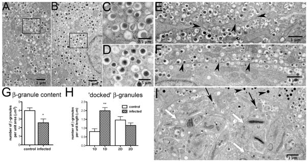
Figure 4. Granule morphology and cellular distribution in control and infected mouse pancreatic islets.
Electron micrographs of T. cruzi-infected or uninfected islets at 30 days post-infection were analyzed for changes in granule number or distribution. During acute infection, amastigotes were observed within the pancreatic β-cells (Figure 4 I, white arrows). However, no parasites were detected in α-cells and the glucagon granules appeared to be intact (Figure 4 I, black arrowheads). The integrity of insulin granules also appeared morphologically normal (Figure 4 A–D, Figure 4 I, black arrows). Compared to tissues from uninfected mice, the total number of insulin granules was reduced in pancreata obtained from mice 30 days post-infection (Figure 4 G). Consistent with decreased 1st phase secretion, in the infected β cells there was a significant increase in the number of docked insulin granules, i.e. granules localized within one granule diameter from the plasma membrane (Nagajyothi et al. 2013). Figure was reprinted with permission of the American Journal of Pathology and Elsevier.

