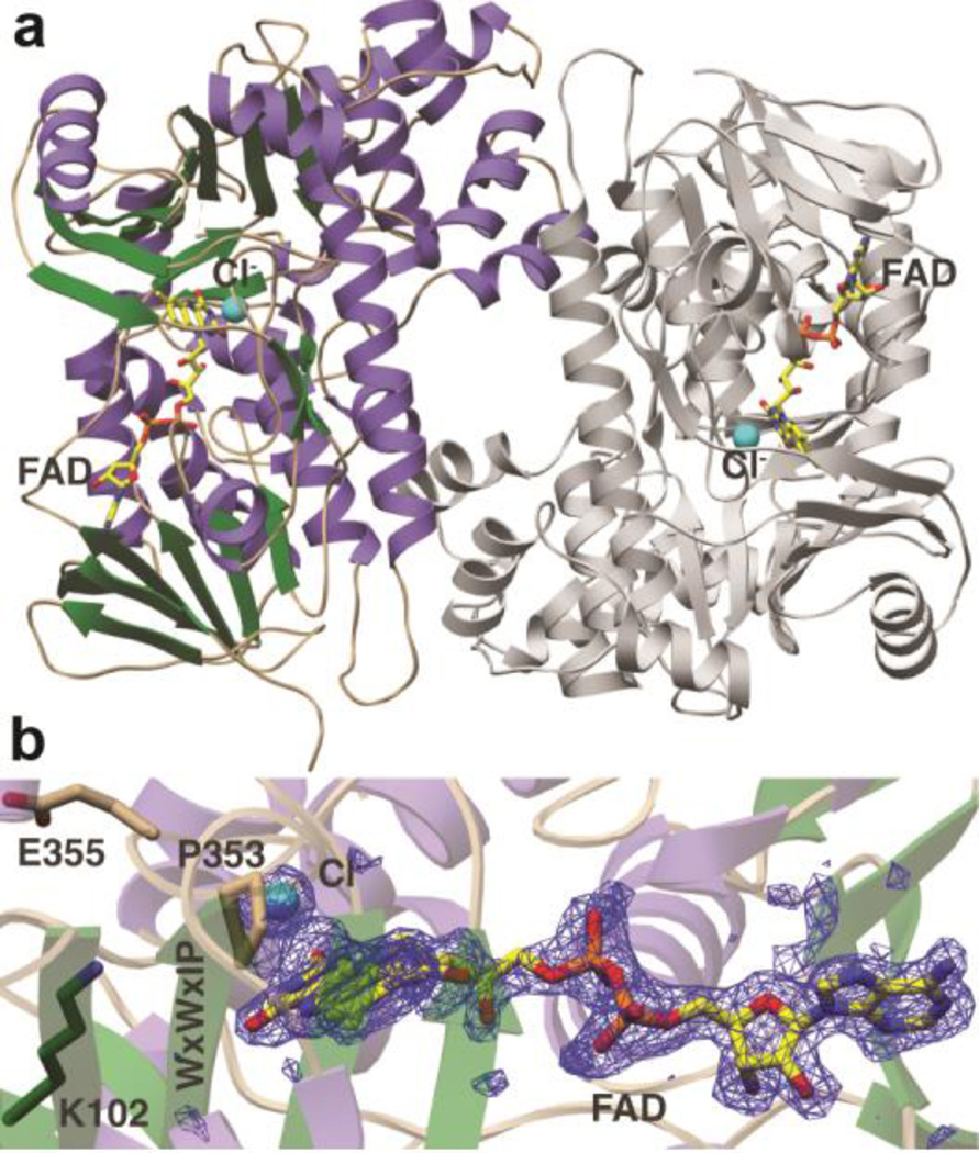
Figure 5.
Crystal structure of the lantibiotic halogenase MibH. (a) Overall cartoon representation of MibH.α helices are shown in purple, β sheets are shown in green, flexible loops are shown in wheat and the second protomer is shown gray. FAD and the Cl− ion are shown as sticks and as a cyan sphere respectively. (b) Simulated annealing omit difference Fourier map (Fo – Fc) contoured to 2.5σ of FAD and Cl−. The conserved Lys/Glu pair involved in chlorination is shown as sticks. The conserved WXWXIP motif is labeled in one amino acid code.

