Fig. 5a–f.
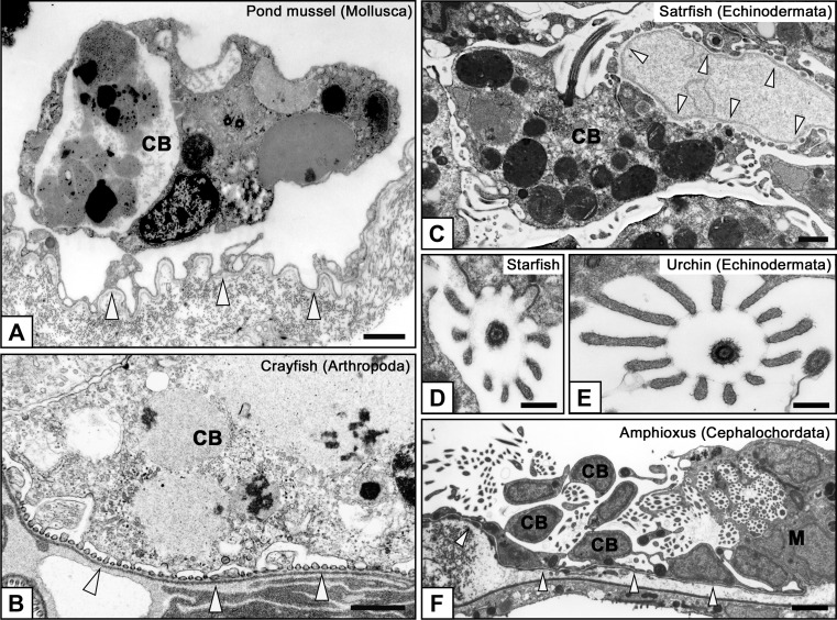
Characteristic structures of podocytes in invertebrates. Well-developed lysosomes are frequently found within the podocyte cell bodies in invertebrates (a–c), suggesting that the podocytes may play a role in the isolation of non-excreted toxic substances to maintain the quality of hemolymph in invertebrates. Podocytes generally possess a single motile cilium on the cell body. In echinoderms and cephalochordates, the motile cilium is protruded together with a microvillus collar, which consists of 10–12 microvilli and surrounds a motile cilium in a circle (d–f). The motile cilium with microvillus collar enters the metanephridium from the nephrostome in cephalochordates and is highly likely to lead the primary urine from the body cavity to metanephridium (f). a Pond mussel, b Crayfish, c, d Starfish, e Sea urchin (Hemicentrotus pulcherrimus), f Amphioxus. Arrowheads basement membrane of podocytes. CB Cell body of podocyte, M metanephridium. Bars a–c 1 μm; d, e 200 nm; f 5 μm

