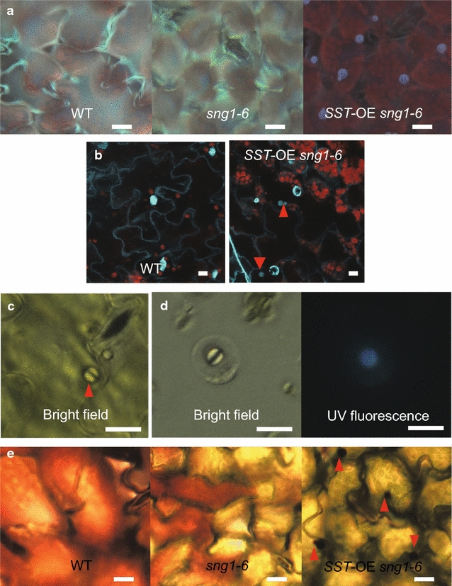
Fig. 6.

Distribution of fluorescent metabolites in 2- to 3-week-old SST-OE sng1-6 plants. a UV fluorescence under microscope. b Presumptive fluorescent particles (arrow heads) after DAPI staining. c Fluorescent particles (arrow heads) in the adaxial epidermis under bright field. d Fluorescent particles in the protoplasts under bright field and UV fluorescence. e Particles (arrow heads) were stained with neutral red. Scale bars 10 μm
