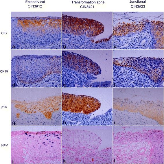
Fig. 4.

Expression pattern of CK7, CK19, p16, and HR HPV in CIN3. HE staining shows representative CIN3s developing in the ectocervix (CIN3#12) (a), transformation zone (CIN3#21) (b), and SCJ (CIN3#23) (c), respectively. CIN3#12 shows patchy staining of CK7 in the upper layer (d), patchy staining of CK19 in the lower layer (g), diffuse staining of p16 (j), mixture of episomal and integrated HPV with remarkable episomal form in the upper layer (m). CIN3#21 and CIN3#23 show diffuse staining of CK7 (e and f), CK19 (h and i), and p16 (k and l). HPV is present in integrated form in CIN3#21 (n) and mixed episomal and integrated form in CIN3#23 (o). Original magnifications: x400
