Fig. 2.

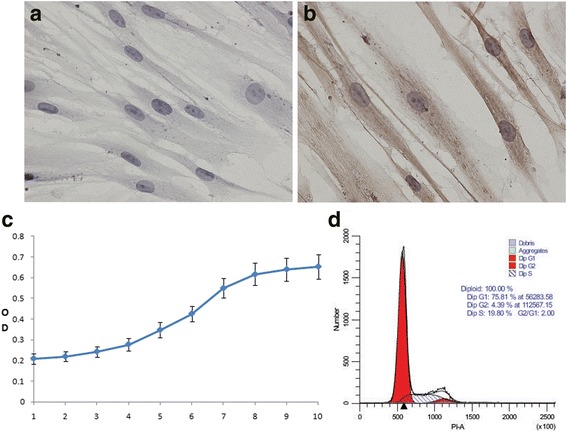
The properties and characteristics of periodontal ligament stem cells. The cells were negative for keratin expression according to immunohistochemical staining (40X) (a). The cells were positive for vimentin expression. Several brown particles in the cytoplasm were observed (40X) (b). Growth was measured at passage 2. The growth curve of PDLSCs exhibited an “S” shape (c). The cell cycle of PDLSCs was examined at passage 5, showing that most cells were in G1 phase; proliferation was slow (d)
