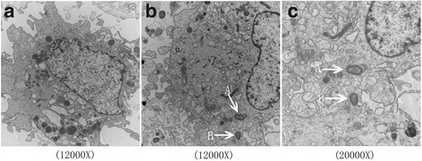
Fig. 6.

PDLSCs infected with P. gingivalis ATCC 33277 under transmission electron microscopy. The nucleus was large and round, and organelles were abundant in PDLSCs (a). P. gingivalis ATCC 33277 could invade PDLSCs after 2 h of incubation (b, c-A). Endocytic vacuoles were not found surrounding internalized P. gingivalis. The bumps observed were stretched membrane where the PDLSCs packaged P. gingivalis ATCC 33277 (b, c-B)
