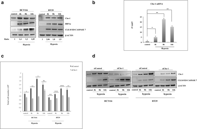
Fig. 1.

Che-1 is involved in the response to hypoxia. a Western blot (WB) analysis performed with the indicated antibodies (Abs) of total cell extracts (TCEs) from HCT116 or HT29 cells exposed to hypoxia (1% O2) for the times indicated. b Quantitative RT–PCR (qRT–PCR) performed in HCT116 cells exposed to hypoxic conditions as in a. Values were normalized to RPL19 expression. Bars represent the standard error of three different experiments. *P ≤ 0,02, **P ≤ 0.002. c HCT116 and HT29 cells were transiently transfected with Stealth siRNA negative control (siControl) or siRNA Che-1 (siChe1) and treated as in a. Cell proliferation was measured by counting the cells daily. A representative histogram is depicted utilizing values from three independent experiments and significance was calculated. *P ≤ 0.02, **P = 0.0005, ***P = 0.0008, ****P ≤ 0,0001, n.s., not significant. d WB analysis with the indicated Abs of TCEs from HCT116 and HT29 cells transiently transfected and treated as in c
