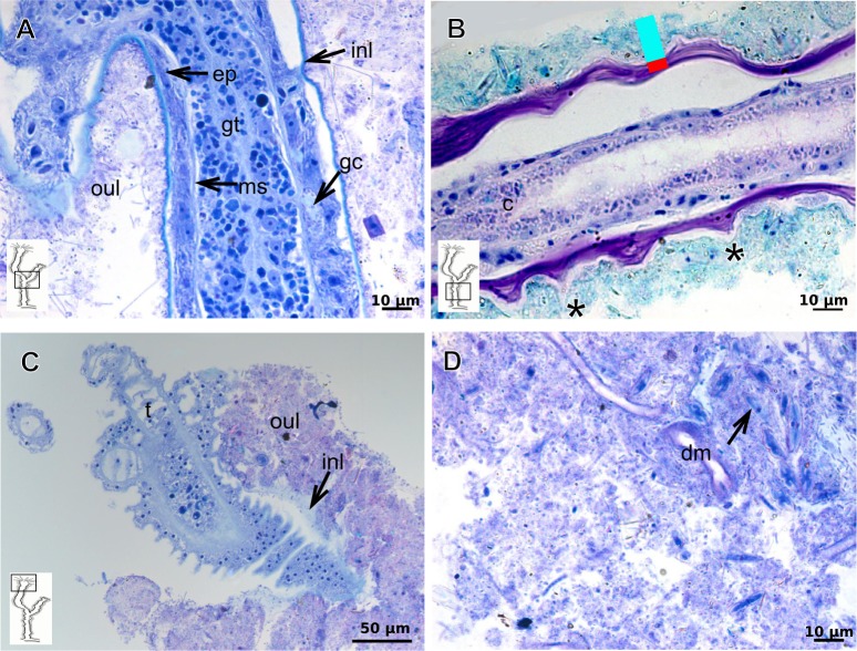
Figure 15. Internal and exoskeletal structure of Leuckartiara cf. octona (Fleming, 1823).
(A) Hydrocaulus and side-branch of the central region of the polyp, stained with TB; (B) hydrocauline exoskeleton, stained with AB + PAS + H; (C)–(D) stained with TB; (C) exoskeleton of the hydranth; (D) outer layer (=exosarc) with organic and inorganic material. Cyan-blue line indicates the outer layer of the exoskeleton, red line indicates the inner layer of the exoskeleton (=perisarc), asterisk indicates “perisarc extensions.” Abbreviations: c, coenosarc; dm, diatoms; ep, epidermis; gc, glandular cells; gt, gastrodermis; inl, inner layer; ms, mesoglea; oul, outer layer; t, tentacle.

