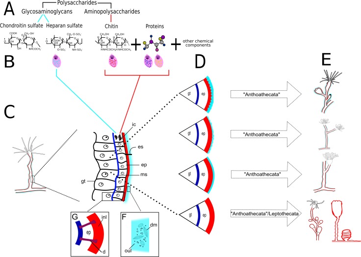
Figure 23. Schematic drawing of different chemical and structural types of the exoskeleton in the Hydroidolina.
(A) Chemical components; (B) scheme of glandular and interstitial cells; (C) coenosarc and exoskeleton; (D) structural types of the exoskeleton; (E) exoskeleton extension in polyps of “Anthoathecata” and Leptothecata; (F) outer layer encrusted with inorganic and organic material; (G) desmocytes connecting inner layer with mesoglea. Cyan-blue line indicates the outer layer of the exoskeleton, red line indicates the inner layer of the exoskeleton. Abbreviations: d, desmocyte; dm, diatoms; ep, epidermis; es, exoskeleton; gt, gastrodermis; ic, interstitial cells; inl, inner layer; ms, mesoglea; oul, outer layer.

