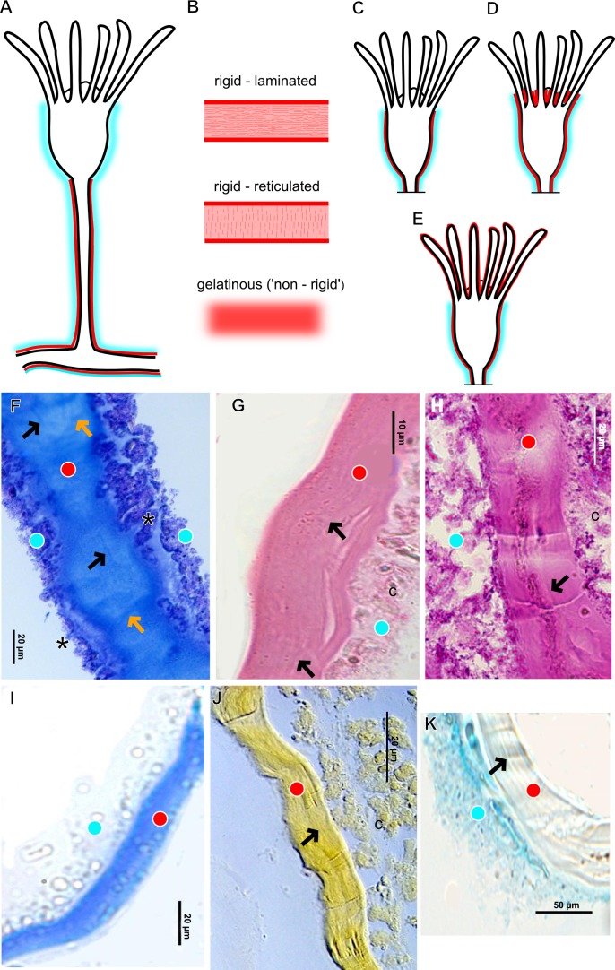
Figure 3. Exoskeletal structure of Bougainvilliidae Lütken, 1850.
(A) Coverage of exoskeletal layers over hydrocaulus; (B) Histological structure of the inner layer (=perisarc) over hydrocaulus; (C)–(E) Coverage of exoskeletal layers over hydranth; (C) reaching the whorl of tentacles; (D) base of the tentacles; (E) inner layer entirely covering the tentacles; (F)–(K) Affinity for chemical tests and details of exoskeleton; (F) Toluidine blue; (G) Eosin; (H) Periodic acid-Schiff; (I) Mercury-bromophenol blue; (J) Naphthol yellow S; (K) Alcian blue pH 2.5. Cyan-blue line and circle indicate the outer layer of the exoskeleton (=exosarc), red line and circle indicate the inner layer of the exoskeleton, black arrow indicates laminae, orange arrow indicates transverse marks, asterisk indicates “perisarc extensions.” Abbreviation: c, coenosarc.

