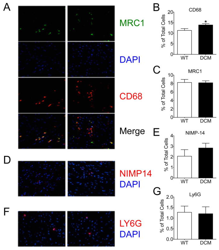
Fig 3. Elevation in CD68+ proinflammatory macrophages in DCM heart tissue.
Representative confocal images and corresponding quantification of (A, B) CD68+ macrophages and (A, C) MRC1+ M2 anti-inflammatory macrophages (n = 58 sections from 5 WT hearts, 75 sections from 5 DCM hearts, *p = 0.016) and their merge, as well as (D, E) NIMP14+ (n = 40 sections from 4 WT hearts, 40 sections from 4 DCM hearts) and (F, G) Ly6G+ (n = 18 sections from 3 WT hearts, 29 sections from 1 DCM heart) neutrophils in WT and DCM heart tissue sections. DAPI (4′,6-diamidino-2-phenylindole).

