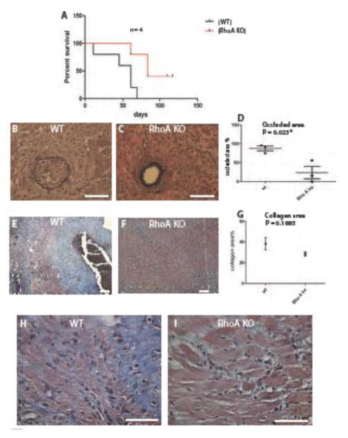
Figure 1. Macrophage specific RhoA deletion inhibits chronic rejection.
(A) Comparison of cardiac allograft survival in RhoAflox/flox (no Cre, labeled wild type, WT) and Lyz2Cre+/−RhoAflox/flox (labeled RhoA KO) recipients. Recipients received 0.25 mg of CTLA4-Ig (administered i.p.) on day 2 and day 4 post- transplantation. (B, C) Sections of transplanted hearts at 50 days post-transplantation stained with VVG. (B) Fully occluded vessel from the RhoAflox/flox (no Cre) recipient and (C) un-occluded vessel from the Lyz2Cre+/− RhoAflox/flox recipient. (D). Graph showing the difference in neointimal inedx between hearts from RhoAflox/flox (no Cre) and Lyz2Cre+/−RhoAflox/flox recipients. Y-axis indicates percentage of occluded area. The difference in vessel occlusion (neointima formation) is statistically significant with P value <0.05. The VVG stained sections were used to calculate neointimal index (vessel occlusion). For comparison of vessel occlusion we inspected 5–8 vessels in each mouse, and 20 vessels were used to calculate neointimal indexes. 3 mice from each group were used. (E, F) Sections of transplanted hearts at 50 days post-transplantation stained with trichrome Masson’s stain. Blue color shows collagen deposition. Heart from RhoAflox/flox (no Cre) recipient (E) shows much more collagen deposition than heart from Lyz2Cre+/−RhoAflox/flox recipient (F). (G) Comparison of collagen deposition between hearts from RhoAflox/flox (no Cre) and Lyz2Cre+/−RhoAflox/flox recipients. Although there is a visible difference in the collagen deposition this difference is statistically insignificant with P value > 0.05. 3 mice from each experimental group were used. (H, I) High magnification of tissue fragments from heart sections shown in E and F illustrates dramatic difference in cardiac muscle tissue integrity. (B, C, H, I) Bar is equal to 50 μm. (E, F) Bar is equal to 100 μm.

