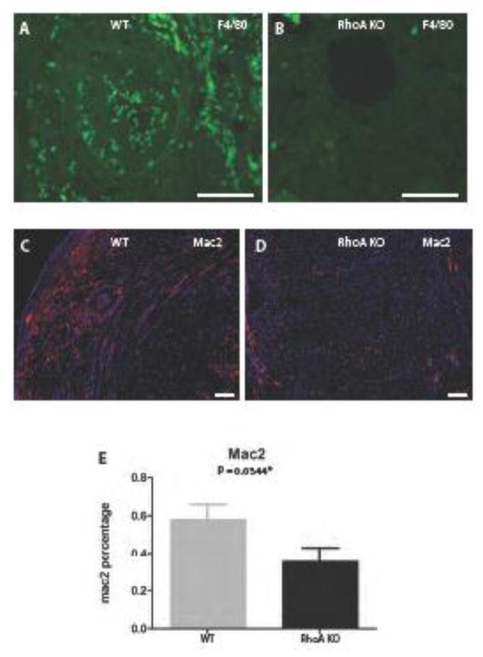
Figure 2. Macrophage specific RhoA deletion inhibits macrophage infiltration of the allograft.

(A, B) Sections of transplanted hearts at 50 days post-transplantation immunostained with antibody against macrophage marker F4/80 conjugated with FITC. (A) High macrophage (green spots) infiltration is present in the heart from the RhoAflox/flox (no Cre; labeled WT) recipient and (B) negligible macrophage infiltration in the heart from Lyz2Cre+/−RhoAflox/flox (labeled RhoA KO) recipient. (C, D) Sections of transplanted hearts at 50 days post-transplantation immunostained with antibody against activated macrophage marker Mac-2 and secondary antibody conjugated with red fluorescein protein. (C) High macrophage (red spots) infiltration is present in the heart from the RhoAflox/flox (no Cre) recipient and (D) low macrophage infiltration in the heart from Lyz2Cre+/−RhoAflox/flox recipient. (E) Graph showing the difference in the Mac-2 positive macrophages between hearts from RhoAflox/flox (no Cre) and Lyz2Cre+/−RhoAflox/flox recipients. The difference is statistically significant with the P value <0.05. 3 mice from each experimental group were used. (A, B) Bar is equal to 50 μm; (C, D) Bar is equal to 100μm.
