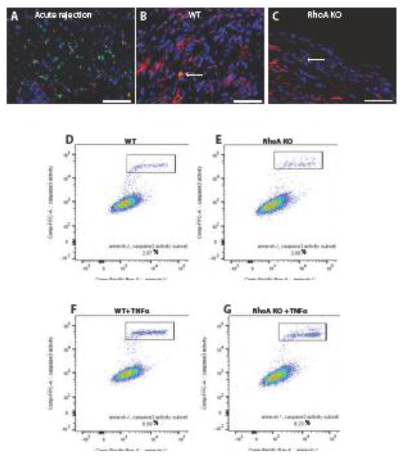
Figure 3. Apoptosis in cardiac allografts and peritoneal macrophages.

(A–C). Sections of hearts fixed at 7 days post-transplantation (acutely rejecting positive control) and at 50 days post-transplantation were stained using Tunel Assay and immunostained with macrophage marker Mac-2 antibody. Nuclei were counterstained with Hoechst. Apoptotic cells are green, macrophages are red and nuclei are blue. (A) Section of the acute rejecting heart at 7 days post-transplantation (positive control) shows high macrophage infiltration (red) and high number of apoptotic cells (green). Hearts from RhoAflox/flox (no Cre; labeled WT) recipient (B) and Lyz2Cre+/−RhoAflox/flox (labeled RhoA KO) recipient (C) show very low number of Tunel positive apoptotic cells. Arrows point to Tunel positive cells. Bar is equal to 50 μm. (D–G) Flow cytometry of apoptosis assay (gating on F4/80+/CD11b+ cells) performed on peritoneal macrophages isolated from RhoAflox/flox (no Cre) (Control) and Lyz2Cre+/−RhoAflox/flox (RhoA KO) mice. (D, E) There is no difference in apoptotic cell percentage between control and RhoA deleted-macrophages. (E, F) RhoA-deleted macrophages are not more vulnerable to the apoptosis-inducing factor (TNFα) than control macrophages. 3 mice from each experimental group were used.
