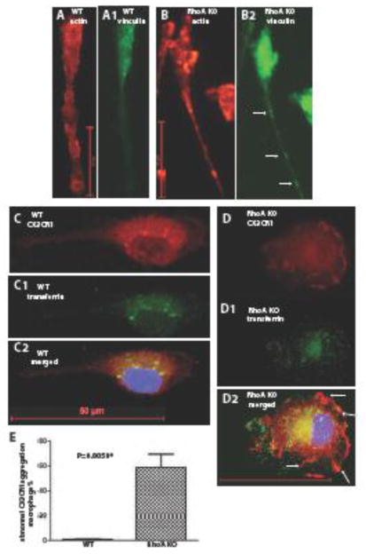
Figure 6. RhoA deletion affects actin cytoskeleton and inhibits association of CX3CR1 with the endososmes.
(A, B) Actin and (A1, B1) anti-vinculin antibody staining of wild type and RhoA deleted bone marrow derived M0 macrophages. RhoA-deleted macrophages show extreme elongation of the tail with visible aggregations of focal adhesions (arrows). (C, D) Wild type and RhoA-deleted bone marrow derived M0 macrophages stained with anti-CX3CR1 antibody and (C1, D1) anti-transferrin receptor (CD71; endosome marker) antibody. (C2, D2) Merged images of CX3CR1 (red), transferrin receptor (green) and nuclear Hoechst staining (blue). In wild type macrophages CX3CR1 co-localized with endosomes, while in RhoA-deleted macrophages the co-localization with endosome is disrupted and CX3CR1 localizes at the macrophage plasma membrane (arrows). Bar is equal to 50 μm.

