
Fig. 3.
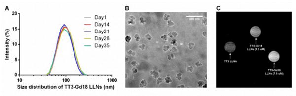
Characterization of TT3-Gd18 LLNs and in vitro MRI. (A) Particle size of TT3-Gd18 LLNs remained constant for over one month. (B) A representative cryo-TEM image of TT3-Gd18 LLNs. Scale bar: 200 nm. (C) A representative T1 weighted image of cell pellets.