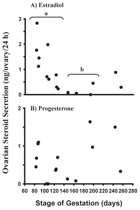
Fig. 3.
Estradiol and progesterone secretion (ng/ovary/24 h) in vitro by ovaries from bovine fetuses at different gestational ages (means ± SEM). Ovaries were cut into pieces (0.5 – 1 mm3). The pieces were then cultured for 24 h and steroid concentrations in the culture medium were determined by RIA. a,b: mean estradiol levels differ (P<0.05). (From Yang & Fortune 2008, with permission.)

