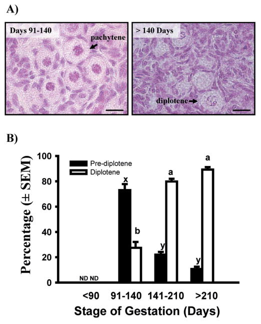
Fig. 7.
A) Photomicrographs showing bovine primordial follicles with oocytes at the pachytene or diplotene stage of first meiotic prophase. Bar = 20 μm B) Percentage of oocytes in primordial follicles at pre-diplotene (leptotene, zygotene, or pachytene) and diplotene stages of first meiotic prophase in bovine fetal ovaries collected at different stages of gestation (means ± SEM). Means with no common letters within meiotic stage across gestational stage (pre-diplotene: x, y; diplotene: a, b) are significantly different (P < 0.05; n = 3–6 fetuses per gestational stage; with 46–207 oocytes in primordial follicles examined per fetus). ND: not detected. (Adapted from Yang & Fortune 2008, with permission.)

