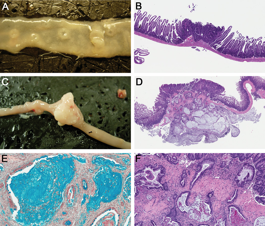
Figure 1. SB-induced intestinal tumor histology.
Representative intestinal (A) adenomas and (C) adenocarcinoma from SB mice. H&E stained sections of representative intestinal (B) adenoma (Magnification, 4X), and (D) mucinous adenocarcinoma (2X). (E) Alcian blue stained mucinous adenocarcinoma (20X). (F) Higher magnification of panel D, (10X), shows neoplastic glands penetrating through the muscularis mucosa.

