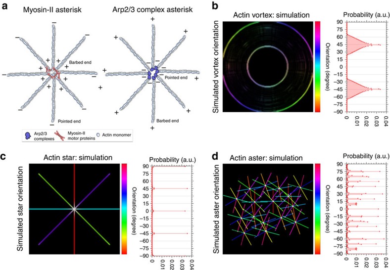
Figure 1. Schematic of the self-organization of the actin cortex.
(a) Two main nucleation pathways of actin patterning: (right) myosin II motor proteins crosslink F-actin (out of actin monomers) at their barbed ends (+) at the pattern centres resulting in the point ends (−) pointing outwards; and (left) Arp2/3 complexes bind to preexisting F-actin and nuclate new filaments from their pointed ends (−) leaving the barbed ends (+) pointing outwards. (b–d) Simulations of self-organizing actin patterns as proposed from in vitro data: vortices (b), asters (c), and stars (d). Left panels: principal spatial organization of actin with colour scale highlighting orientation of F-actin arms in space (light blue horizontal to red vertical, −90 to 90). Right panels: Frequency histogram of spatial orientations of F-actin arms (given as probability distribution), highlighting characteristic distributions for each case.

