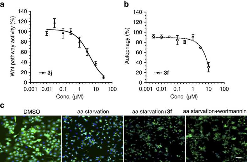
Figure 8. Influence of representative compounds on Wnt signalling (3j) and autophagy (3f).
(a) Dose-dependent inhibition of the Wnt pathway as determined by means of Wnt reporter gene. HEK293 cells stably transfected with the human Frizzled-1 receptor and a TOPFLASH-driven luciferase reporter gene were treated with different concentrations of 3j for 6 h. Expression of the firefly luciferase as a reporter gene was the determined by means of luminescence as readout. Nonlinear regression analysis was performed using a four parameter fit. Data are mean values of three independent experiments (n=3)±s.d. (b) Dose–response curve for inhibition of autophagy by 3f. MCF7-GFP-LC3 cells were deprived of amino acids to induce autophagy and treated with the different concentration of 3f for 3 h. GFP-LC3 was detected as a measure of autophagosome formation. Nonlinear regression analysis was performed using a four parameter fit. Data are mean values of three independent experiments (n=3)±s.d. (c) MCF7 cells that stably express GFP-LC3 (green) were starved for amino acids (aa) in the presence of the compound 3f (10 μM) or Wortmannin (3 μM) for 3 h before fixation and staining of the DNA using Hoechst 33342 (blue). Autophagy induction is detected as an accumulation of GFP-LC3 puncta on starvation. Scale bar, 10 μm.

