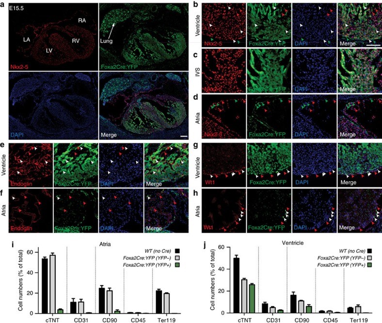
Figure 5. Contribution of Foxa2-vCPs to the major cardiovascular lineages.
(a) Heart sections from E15.5 Foxa2Cre:YFP embryos stained with antibodies against YFP and Nkx2–5 to label cardiomyocytes. Tile scan image showing heart and lung. (b–h) IF analysis of E15.5 Foxa2Cre:YFP embryos using antibodies against YFP and Nkx2–5 (myocardium, b–d), Endoglin (endocardium, e,f) or Wt1 (epicardium, g,h). Detailed areas of ventricle (b,e,g), interventricular septum (c) and atria (d,f,h) are shown. Arrowheads indicate cells that express the relevant lineage markers alone (red) or that co-express YFP and the relevant lineage marker (white). Images are representative examples from multiple experiments. (i,j) Flow cytometry analysis of dissociated cells of atrial (i) and ventricular (j) chambers of E13.5 Foxa2Cre:YFP hearts with antibodies against cTnT (cardiomyocytes), CD31 (endothelial), CD90 (mesenchymal, haematopoietic, fibroblast, epicardium), CD45 (leukocyte) and Ter119 (erythroid). Data are mean±s.e.m. of n=7 hearts. Scale bars, 100 μm. LA, left atria; LV, left ventricle; RA, right atria; RV, right ventricle.

