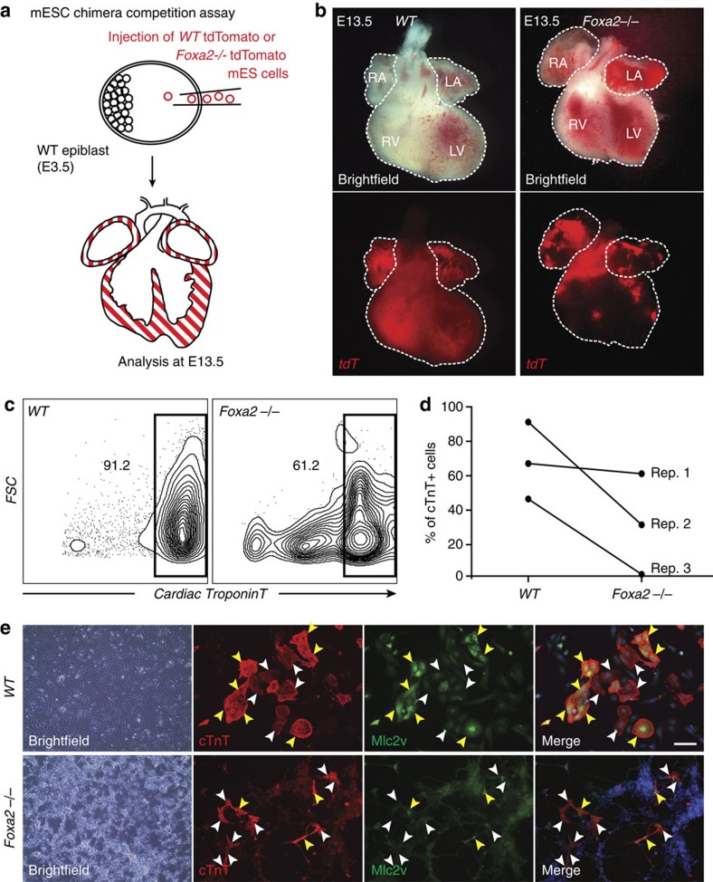
Figure 7. Foxa2 is necessary for the generation of ventricular cells during cardiac development.
(a) Schematic of mESC chimera competition assay. Fluorescently labelled WT or Foxa2−/− mESCs are injected into unlabelled WT blastocysts at E3.5. Embryos are collected at E13.5 and the distribution of tdT+ cells is observed. (b) Whole-mount imaging of typical WT (left) and Foxa2−/− (right) mESC-injected hearts at E13.5. (c) Flow cytometry analysis of cell populations differentiated from WT and Foxa2−/− mESC cells. Cells at day 10 of differentiation were dissociated and analysed by flow cytometry for the cardiac marker cTnT. (d) Quantification of c. Paired data are plotted for n=3 replicates. (e) Cells at day 10 of differentiation from WT and Foxa2−/− mESCs were plated and IF analysis was performed with antibodies against cTnT (red) and the ventricular-specific marker Mlc2v (green). Yellow arrowheads illustrate overlap of cTnT and Mlc2v. White arrowheads indicate cTnT cells that are not stained for Mlc2v. Scale bar, 50 μm. LA, left atria; LV, left ventricle; RA, right atria; RV, right ventricle.

