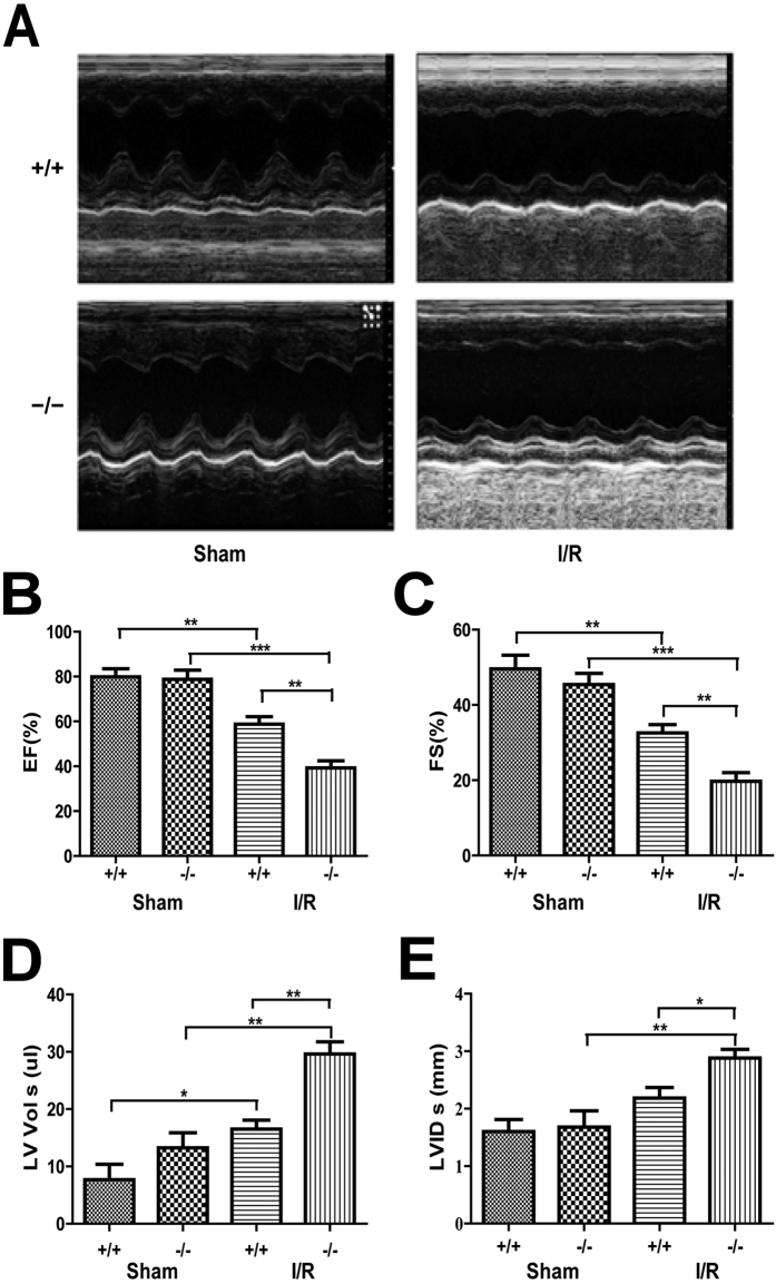
Figure 2. Plin5 deficiency aggravates heart dysfunction following I/R injury.
(A) Echocardiography was performed at the end of reperfusion, and representative M-mode echocardiograms were recorded in all groups. Mice without LAD occlusion served as basal controls (Sham group). (n = 6). (B–E) Cardiac function was examined by echocardiography after I/R surgery. The EF (B) and FS (C) values of Plin5-null mice decreased more significantly than those in wild-type mice after I/R surgery, whereas LV Vol;s (D) and LVID;s (E) also increased significantly in Plin5-null mice after I/R surgery. (n = 6). The columns and errors bars represent means ± SEM. *P < 0.05; **P < 0.01; ***P < 0.001. EF, ejection fraction; FS, fractional shortening; LV Vol;s, left ventricular contraction volume; LVID;s, left ventricular internal diameter at end-systole.

