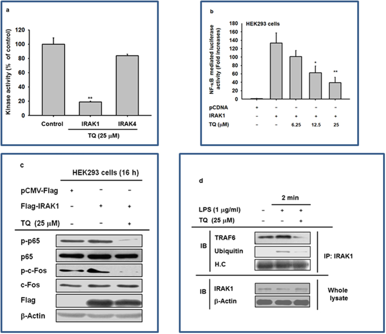
Figure 5. Effect of TQ on the activation of IRAK1.
(a) Kinase activities of IRAK1 and IRAK4 were determined by a kinase profiler service using purified enzymes and substrate. The vehicle control was set to 100% activity for IRAK1 and IRAK4 enzymes for the purpose of comparison with treated cells. (b) Effect of TQ on IRAK1-induced NF-κB activation was measured by a reporter gene assay. The luciferase activity of HEK293 cells transfected with NF-κB-Luc (1 μg/mL) and IRAK1 in the presence or absence of TQ was measured using a luminometer. (c) Effect of TQ on the activation of AP-1 and NF-κB upon IRAK1 overexpression was assessed by immunoblot analysis of phosphorylated and total levels of p65 and c-Jun. (d) Effect of TQ on the formation of the IRAK1-TRAF6 complex and ubiquitinylation was determined by immunoblot analysis. The blots in c and d were obtained under the same experimental conditions and are shown as cropped blots (original blots with indicated cropping lines are shown in Supplementary Figure S1). The data shown represent mean ± SD of three (a) or five (b) samples. Results in c and d are shown for one representative experiment of three. *p < 0.05 and **p < 0.01 compared with control.

