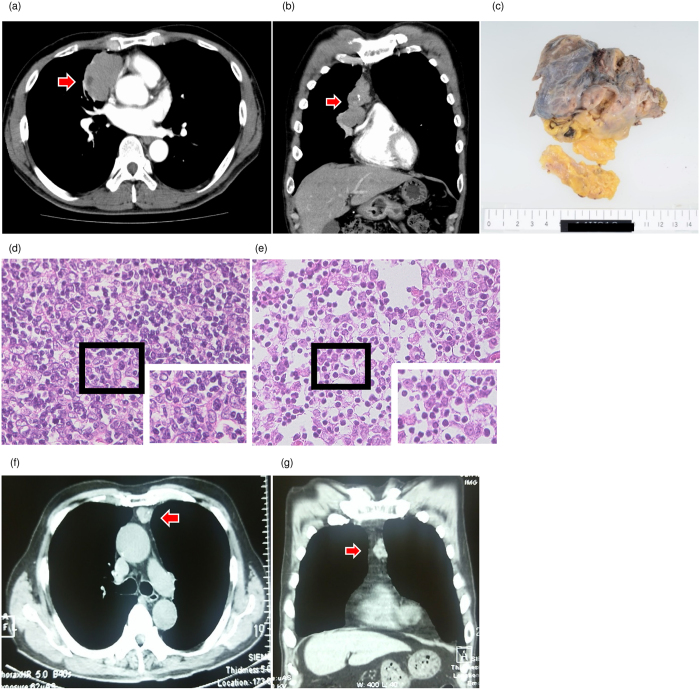
Figure 1. The findings of thymomas.
(a,b) Computed tomography (CT) findings of thymomas of Patient 1. (a) Horizontal plane, (b) Coronal plane. Arrows indicate thymoma. (c) Macroscopic finding of the thymoma in patient 1. (d,e) Hematoxylin and eosin stain of thymoma tissues. (d) Patient 1, (e) Patient 2 (×400). (f,g) CT findings of the thymoma of patient 3. (f) Horizontal plane, (g) Coronal plane. Arrows indicate thymoma.

